


Radiology of the PleuraRadiology of the Pleura
Adam Guttentag M.D.Adam Guttentag M.D.
Pleural diseasesPleural diseases
EffusionEffusion
SimpleSimple
Complex / loculatedComplex / loculated
Pleural massesPleural masses
Pleural plaquesPleural plaques
Primary massesPrimary masses
Metastatic diseaseMetastatic disease
Pleural EffusionRadiographic evaluationPleural EffusionRadiographic evaluation
Recognizing an effusionRecognizing an effusion
Size: (“can I tap it?”)Size: (“can I tap it?”)
Simple layering with gravity vs. loculationsSimple layering with gravity vs. loculations
CT vs. CXRCT vs. CXR
Pleural EffusionPleural Effusion
Radiographic signs :Radiographic signs :
Learn to recognize them!Learn to recognize them!
Meniscus signMeniscus sign
may not be present onrecumbent or semi-erect filmmay not be present onrecumbent or semi-erect film

Pleural EffusionPleural Effusion
Earliest sign on frontal film is laminareffusion or blunting of CP angle.Earliest sign on frontal film is laminareffusion or blunting of CP angle.
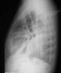
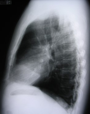
Lateral film is more sensitive thanfrontal film:Lateral film is more sensitive thanfrontal film:
Posterior CP angle is more inferior thanlateral one.Posterior CP angle is more inferior thanlateral one.
Very small effusions can be seen on theCXR (<20cc).Very small effusions can be seen on theCXR (<20cc).
Pleural EffusionPleural Effusion
Radiographic signs:Radiographic signs:
Meniscus signMeniscus sign
Laminar effusionLaminar effusion
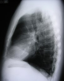
Laminar Effusion
Visceralpleura
Parietalpleura
Blunted posterior CP angle
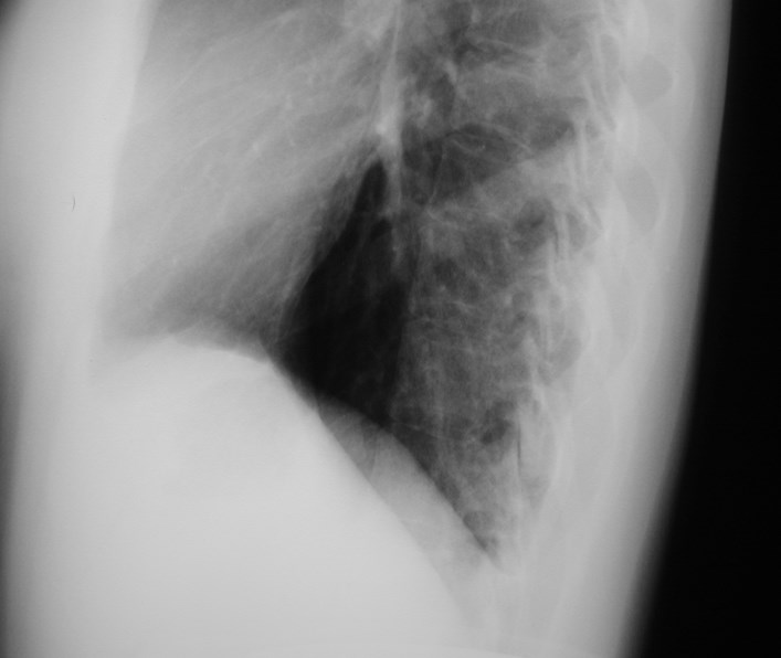
Diaphragm projected here on film
Bottom of pleural space is here
Remember that the lateral film is moresensitive for finding effusions!
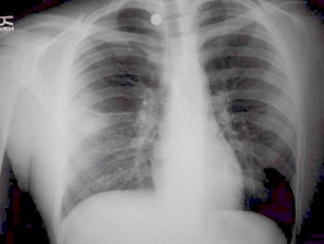
Pleural effusionPleural effusion
Radiographic signs - learn torecognize it!Radiographic signs - learn torecognize it!
Meniscus signMeniscus sign
may not be present onrecumbent filmmay not be present onrecumbent film
Laminar effusionLaminar effusion
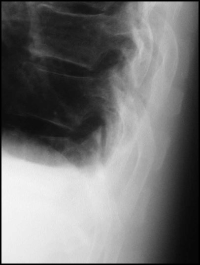
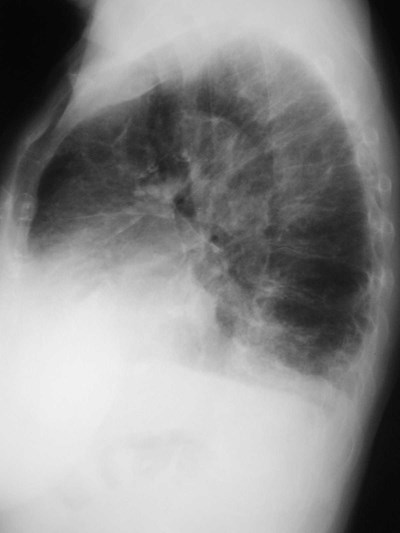
Fluid in a fissureFluid in a fissure
Fluid entering a fissure
“Pseudotumor”
2 weeks earlier
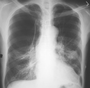
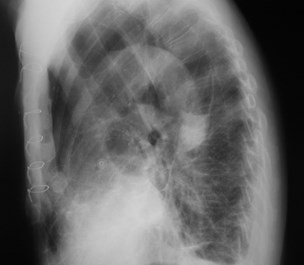
3 days later
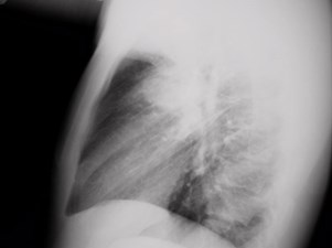
Pleural EffusionPleural Effusion
Radiographic signs - learn torecognize it!Radiographic signs - learn torecognize it!
Meniscus signMeniscus sign
may not be present onrecumbent filmmay not be present onrecumbent film
Laminar effusionLaminar effusion
Fluid in fissureFluid in fissure
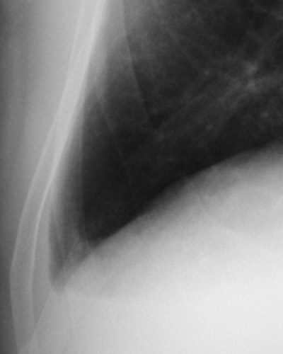
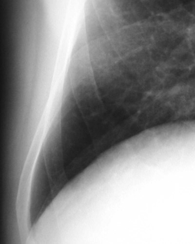
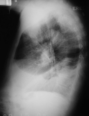
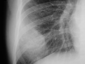
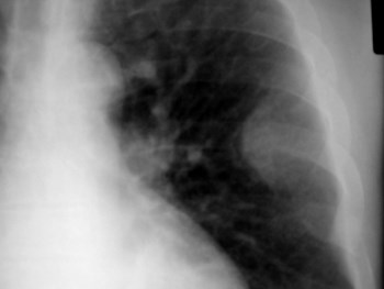
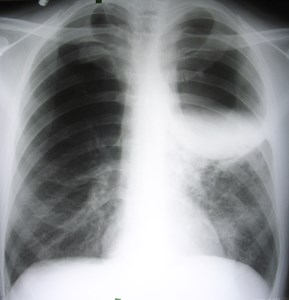
Subpulmonic effusionSubpulmonic effusion
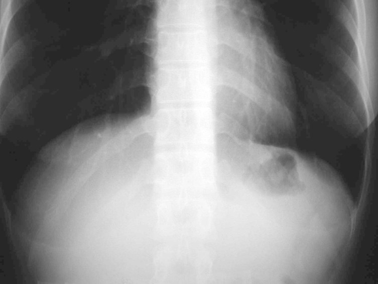
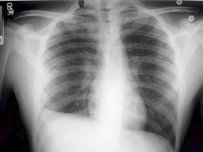
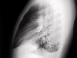
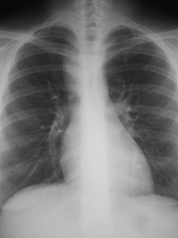
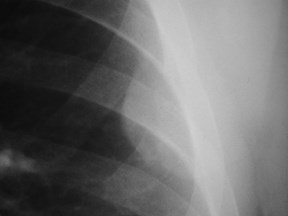
Subpulmonic EffusionSubpulmonic Effusion
Lung floats on effusionLung floats on effusion
Lateral peaking of“diaphragm”Lateral peaking of“diaphragm”
Change in “diaphragm”contourChange in “diaphragm”contour
Distance to stomachbubble - upright view onlyDistance to stomachbubble - upright view only

Subpulmonic EffusionSubpulmonic Effusion
Distance to stomach bubble shouldbe less than 2 cm.
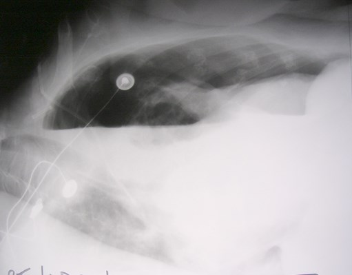
Lateral Decubitus FilmLateral Decubitus Film
Frontal view with patient lying on his sideFrontal view with patient lying on his side
Cross table lateral techniqueCross table lateral technique
Non-dependent lung is hyperinflatedNon-dependent lung is hyperinflated
Free fluid will layer along dependent chest wallFree fluid will layer along dependent chest wall
Quantify (?) effusionQuantify (?) effusion
Simple or loculated?Simple or loculated?
Bonus is good look at “up” lung and findingunexpected contralateral effusionBonus is good look at “up” lung and findingunexpected contralateral effusion
Get both decub’s!Get both decub’s!
Fluid may change its appearance dailyFluid may change its appearance daily
Rapid change in appearance ofpleural effusionRapid change in appearance ofpleural effusion
erect
supine
next day
Simple vs Complex EffusionSimple vs Complex Effusion
Exudate, transudate, chylousExudate, transudate, chylous
Defined by lab dataDefined by lab data
pH, glucose, protein, cell count and differentialpH, glucose, protein, cell count and differential
Not always good correlation with radiographic appearance at CXRand CTNot always good correlation with radiographic appearance at CXRand CT
Simple:Simple:
free layering, no pleural thickeningfree layering, no pleural thickening
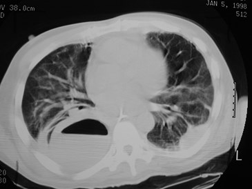
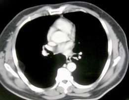
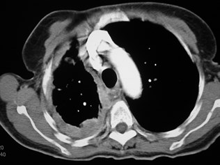
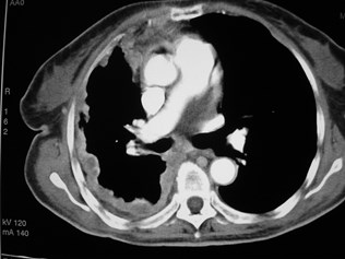
Empyema:Empyema:
often loculated, with smoothly thickened, enhancing pleuraoften loculated, with smoothly thickened, enhancing pleura
May have air within it, multiple loculationsMay have air within it, multiple loculations
CT often reveals unexpected effusion and loculationCT often reveals unexpected effusion and loculation
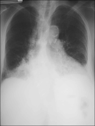
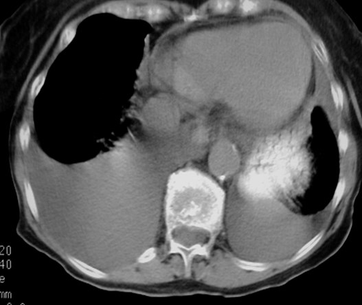
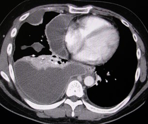
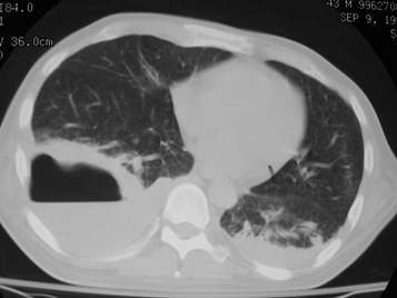
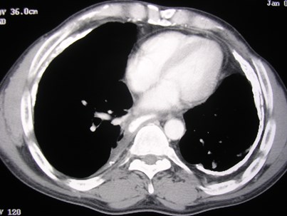
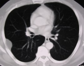
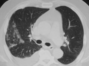
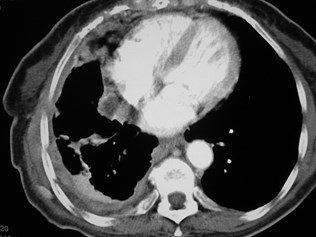
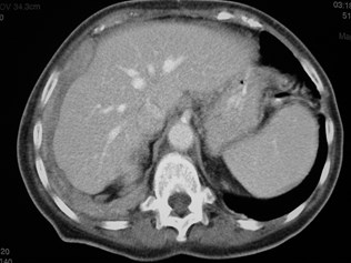
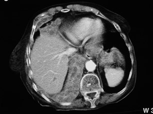
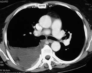
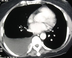
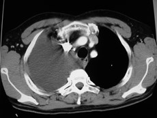
Complex EffusionComplex Effusion
Not all simple appearing effusions arereally simple!
Metasatic breast Cawith trapped lung
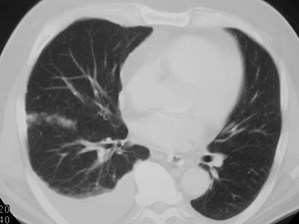
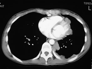
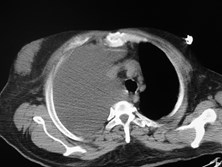
EmpyemaEmpyema
“split pleura” sign
EmpyemaEmpyema
Pleural MassesPleural Masses
PrimaryPrimary
Asbestos related pleural plaquesAsbestos related pleural plaques
Pleural “cloak”Pleural “cloak”
(Subpleural masses eg lipoma, rib lesions)(Subpleural masses eg lipoma, rib lesions)
Localized fibrous tumor of the pleuraLocalized fibrous tumor of the pleura
Malignant mesotheliomaMalignant mesothelioma
MetastaticMetastatic
LungLung
BreastBreast
OvarianOvarian
LymphomaLymphoma

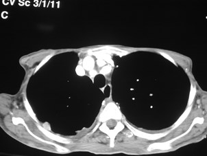
Asbestos related pleuralplaquesAsbestos related pleuralplaques
Fibrous plaques associatedwith asbestos exposureFibrous plaques associatedwith asbestos exposure
Not “asbestosis”!Not “asbestosis”!
Clinically insignificantClinically insignificant
Parietal pleuraParietal pleura
BilateralBilateral
+/- Calcified+/- Calcified
Often involve diaphragmOften involve diaphragm
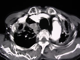
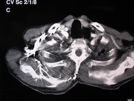
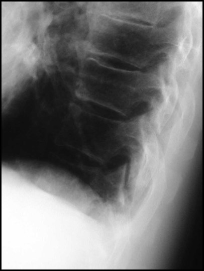
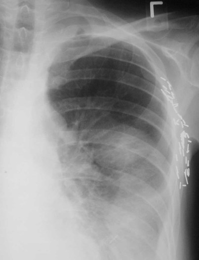
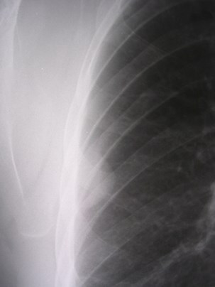
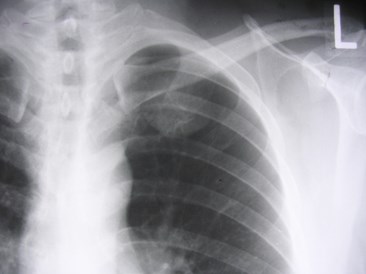
Pleural “cloak”Pleural “cloak”
Usually unilateralUsually unilateral
Broad and thickBroad and thick
Displaced inwardfrom chest wallDisplaced inwardfrom chest wall
Related to priorpleural infection orhemorrhageRelated to priorpleural infection orhemorrhage
TB empyema mostcommon causeTB empyema mostcommon cause
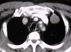
Bilateral Pleural CloakBilateral Pleural Cloak
Pleural massesPleural masses
Radiographic signs:Radiographic signs:
Smooth inner borderSmooth inner border
Poorly visualized outerborderPoorly visualized outerborder
Within a fissureWithin a fissure
Know normal locationsof fissuresKnow normal locationsof fissures
Look for signs of rib involvement!Makes mass most likely chest wall in originLook for signs of rib involvement!Makes mass most likely chest wall in origin
Subpleural LipomaSubpleural Lipoma
Localized Fibrous Tumor of the PleuraLocalized Fibrous Tumor of the Pleura
Localized Fibrous Tumor of the PleuraLocalized Fibrous Tumor of the Pleura
Rare usually benign neoplasmRare usually benign neoplasm
NOT “benign mesothelioma” or “pleural fibroma”NOT “benign mesothelioma” or “pleural fibroma”
Unrelated to smoking or asbestosUnrelated to smoking or asbestos
Sx uncommon unless largeSx uncommon unless large
Cough, chest pain, dyspneaCough, chest pain, dyspnea
HPOA, clubbing, hypoglycemiaHPOA, clubbing, hypoglycemia
Majority attached to visceral pleura, may be infissure, 50% pedunculatedMajority attached to visceral pleura, may be infissure, 50% pedunculated
Effusion in 20%Effusion in 20%
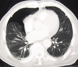
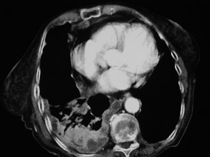
Metastatic Nodules in FissuresMetastatic Nodules in Fissures
Malignant MesotheliomaMalignant Mesothelioma
Metastatic Breast CaMetastatic Breast Ca
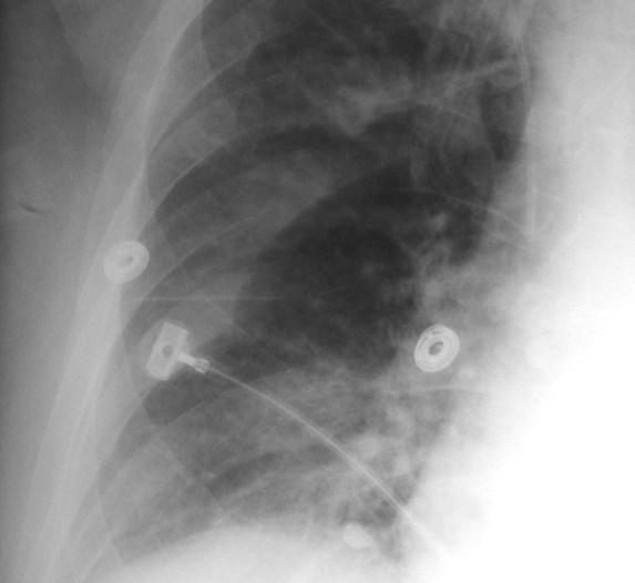
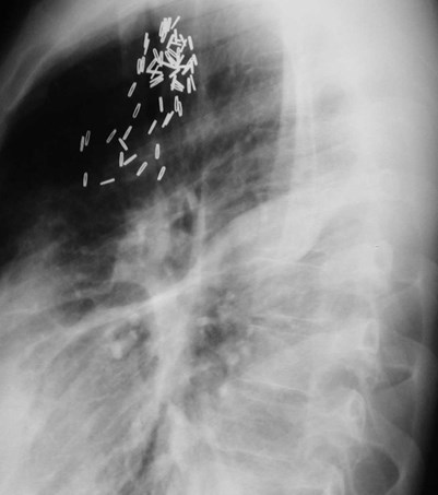
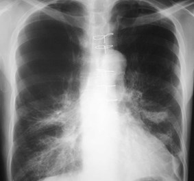
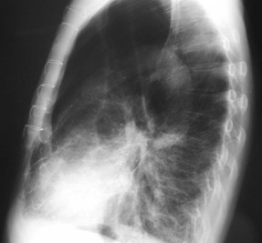
Talc PleurodesisTalc Pleurodesis

Talc PleurodesisTalc Pleurodesis
Aim to produce adhesionbetween visceral and parietalpleuraAim to produce adhesionbetween visceral and parietalpleura
Management of benign andmalignant pleural effusionsManagement of benign andmalignant pleural effusions
Prevention of recurrentpneumothoraxPrevention of recurrentpneumothorax
Closure of bronchopleural fistulaClosure of bronchopleural fistula
Beware positive PET scanafterwards!Beware positive PET scanafterwards!
Malignant vs. Benign PleuralThickening?Malignant vs. Benign PleuralThickening?
Favoring malignant:Favoring malignant:
Areas > 2 cm thickAreas > 2 cm thick
Irregular, nodular massesIrregular, nodular masses
Invasion of chest wallInvasion of chest wall
Mediastinal surface involvementMediastinal surface involvement
Favoring benign:Favoring benign:
CalcificationCalcification
Smooth, < 1 cm thickSmooth, < 1 cm thick
Mesothelioma
Empyema
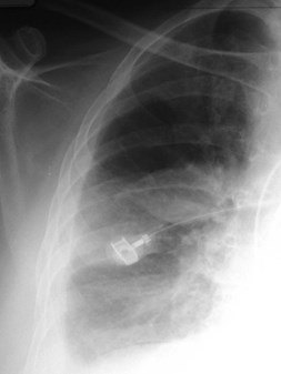
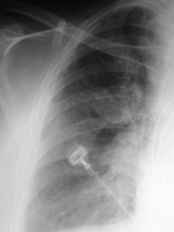
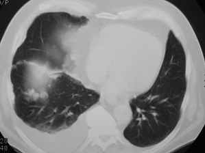
Malignant Pleural NodulesMalignant Pleural Nodules
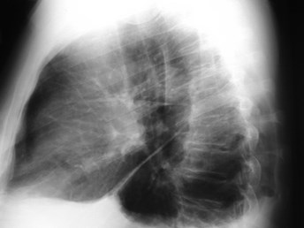
Empyema or Lung Abscess?Empyema or Lung Abscess?
Empyema:Empyema:
Lenticular shapeLenticular shape
Air fluid level different lengths on PA and lateralfilms.Air fluid level different lengths on PA and lateralfilms.
Air fluid level across entire hemithorax = pleuralAir fluid level across entire hemithorax = pleural
Abscess:Abscess:
More spherical shapeMore spherical shape
Air fluid level more equal in length on PA andlateral filmsAir fluid level more equal in length on PA andlateral films
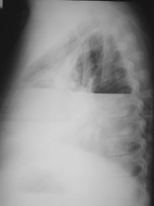
Air fluid level across entirethoraxAir fluid level across entirethorax
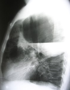
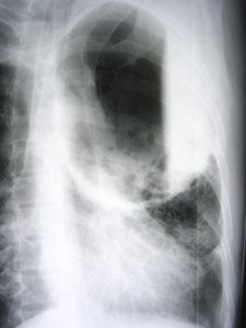
Lung or pleuralcollection?
The old filmgives theanswer!
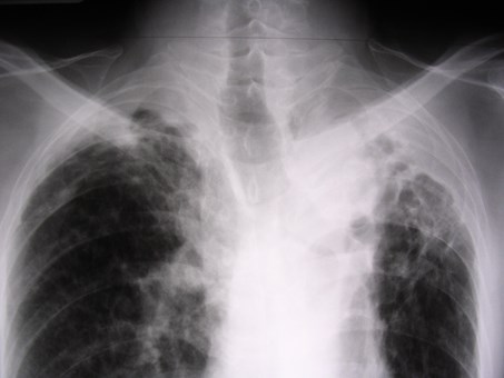
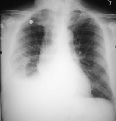
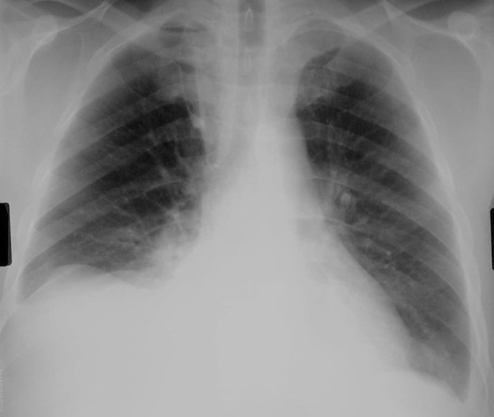
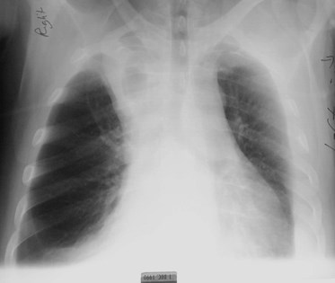
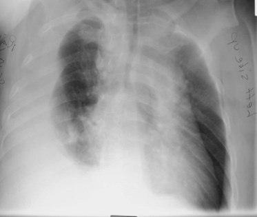
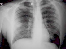
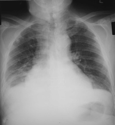
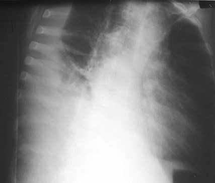
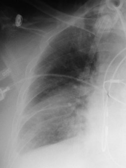
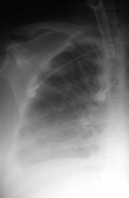
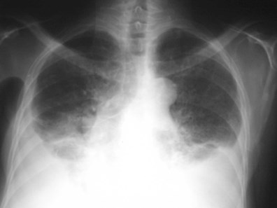
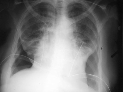
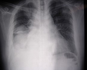
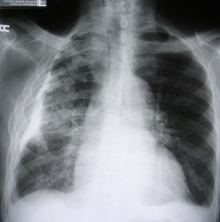
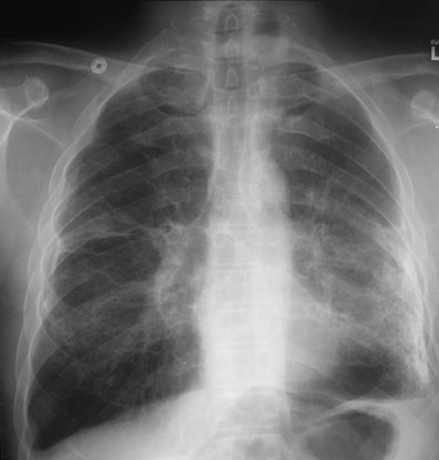
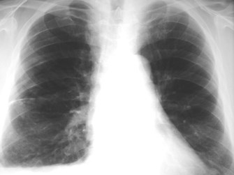
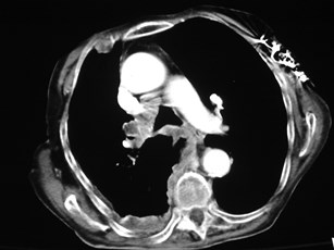
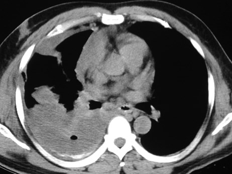
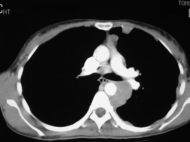
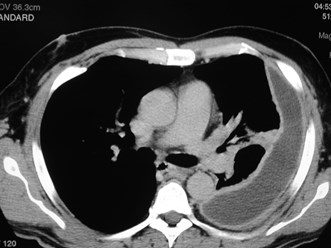
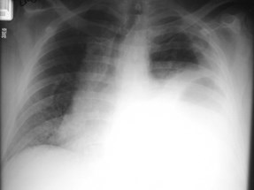
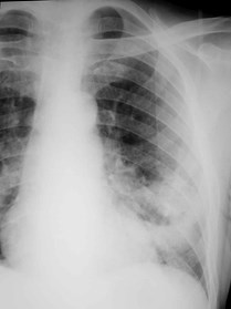
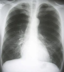
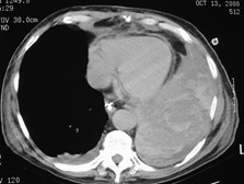
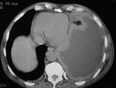
Special Case: Effusion fills most of hemithoraxSpecial Case: Effusion fills most of hemithorax
MalignantMalignant
TBTB
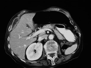
Hepatic effusionHepatic effusion
PancreatitisPancreatitis
HematomaHematoma
ChylothoraxChylothorax
mesothelioma
cirrhosis
TB
hemorrhage
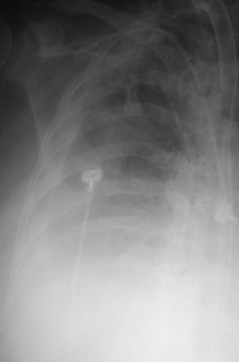
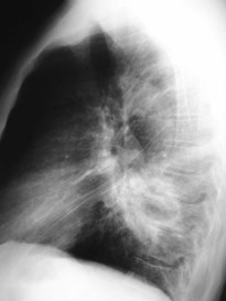
Apical Pleural Thickening?Apical Pleural Thickening?